2024/09/24

Analog Dimmer Switch Points to Intelligent Design and Creation

H3K9 methylation is more like a "dimmer switch" that fine-tunes DNA transcription


https://phys.org/news/2024-06-dna-transcription-paradoxically.html

Excerpt: "Researchers led by Kannosuke Yabe, Asuka Kamio, and Soichi Inagaki of the University of Tokyo have discovered that in thale cresses histone H3 lysine-9 (H3K9) methylation, conventionally thought to be a mark of turning off gene transcription, can also turn on gene expression via the interactions of two other proteins and histone marks.

The molecular mechanisms demonstrate that rather than functioning as a simple "off switch," H3K9 methylation is more like a "dimmer switch" that fine-tunes DNA transcription. The discovery suggests there might be similar mechanisms in other organisms, too. The findings were published in the journal Science Advances.

DNA is often called the "blueprint of biological organisms." However, calling it the "toolbox of cells" might be more accurate because cells also need to control which genes, the basic building blocks of DNA, are transcribed, or in other words, "turned on or off."


This is epigenetics, and it involves the complex interactions of many types of proteins, such as histones. H3K9 methylation is an epigenetic mark associated with turning off DNA transcription. Even though H3K9 methylation was discovered 25 years ago, not all of its molecular mechanisms have been clarified.

"Biological systems are so complex," says Inagaki, the principal investigator, "that it is almost impossible for us to understand exactly how life works. But we can try to understand a tiny part of it. The regulation of gene activity is fundamental to life and is connected to a lot of biological phenomena."

The researchers chose to investigate the molecular mechanisms of gene regulation in Arabidopsis thaliana, commonly known as thale cress. The team used a technique called chromatin immunoprecipitation sequencing (ChIP-seq). This technique provides a detailed view of how proteins interact with DNA. It can be used to analyze the locations of protein modifications very precisely, making it a befitting tool to investigate histone methylation. Then, the results of H3K9 methylation's peculiar role came in.

"At first, I did not pay attention to the results of the analysis," Inagaki remembers, "and did not do any further research on the subject for about a year. I overlooked the finding because it was so unexpected. But one day I had a eureka moment and everything made sense in my head. After that, proving the hypothesis that H3K9 methylation had a dual role went smoothly."

H3K9 methylation's dual role is achieved via two other proteins, LDL2 and ASHH3. LDL2 helps to turn off genes by removing another histone mark, H3K4 methylation. ASHH3 turns the gene on by stopping LDL2 from working via a third histone mark, H3K36 methylation. The complex relationship of the three histone marks (H3K9, H3K4, H3K36) determines the gene's activity.

"I'm happy that we discovered the fundamental aspect of gene regulation by H3K9 methylation, even though many studies around the function and controlling mechanisms of H3K9 methylation have been conducted in many organisms. I hope that this finding will stimulate further scientific endeavors to elucidate how gene regulation works," Inagaki says."

My comment: "Three epigenetic markers (histone epigenetic markings) are almost atom-level digital information units. Their relationship and how they combined affect the DNA transcription activity and strength. This means that digital information is converted to analog information. This kind of information usage points to a brilliant information handling mechanism, Intelligent Design and Creation. Evolution never happened."

2024/09/22

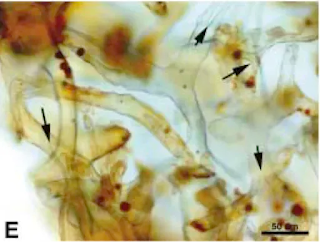
Red Blood Cells in T Rex Soft Tissue Samples Contain C14 Isotope

A red blood cell must be relatively recent and contain C-14 in proportions similar to those in the living environment

A red blood cell (RBC) that has lost all of its carbon-14 (C-14) would be far too old to retain its red color. Here's why:

Key Points:

  1. C-14 Decay and Timeframe:

    • C-14 has a half-life of about 5,730 years. After about 50,000 to 60,000 years, C-14 levels drop to nearly undetectable amounts. If an RBC has lost all of its C-14, it would mean that it is tens of thousands of years old.

  2. Red Blood Cell Decomposition:

    • Red blood cells are made of organic materials that degrade relatively quickly after death. In a living organism, RBCs are constantly replaced and maintained. Once the organism dies or the cells are isolated, they start breaking down rapidly.
    • The red color of RBCs comes from hemoglobin, which degrades and loses its red color relatively quickly (within days to weeks) after cell death due to oxidation and other biochemical changes.
  3. Preservation and Color Retention:

    • To retain its red color, a blood cell would need to be extremely well-preserved, typically through special conditions such as freezing, rapid desiccation, or encapsulation in a way that prevents oxidation and degradation. Rapid burial under mud layers explains this kind of encapsulation. These observations give support to the Biblical flood.

Conclusion:

A blood cell that has lost all its C-14 (indicating an age of over 50,000 years) would not still be red. The organic components would have long since broken down, and the red color would have faded. If a blood cell is still visibly red, it must be relatively recent and contain C-14 in proportions similar to those in the living environment. If the samples contain a C14 isotope, then they can't be millions of years old.


2024/09/20

Change in Allele Frequency is not evidence for Evolution

Change in Allele Frequency is Mostly Due to C>T Genetic Errors: A Mechanism of Rapid Genomic Decline


Introduction

Genomic stability is crucial for the survival and proper functioning of organisms. However, mutations can compromise the genome's integrity, leading to various consequences ranging from minor defects to species extinction. One of the most prevalent mutation types in the human genome is the C>T substitution, primarily resulting from the deamination of methylated cytosines at CpG sites. This mutation type has profound effects on the genomic structure, functioning, and long-term survival of species. In this article, we explore how C>T mutations contribute to a shift in allele frequencies, how they affect genomic stability, and the potential long-term biological consequences. Ultimately, this pattern of mutation may explain rapid genomic decline and even species extinction over relatively short time scales.

Mechanisms of C>T Mutations and Their Impact on Genome Integrity

The C>T mutation occurs when cytosine (C) in the DNA is methylated to form 5-methylcytosine. Over time, this methylated cytosine is prone to spontaneous deamination, resulting in its conversion to thymine (T). CpG sites—locations where cytosine is adjacent to guanine—are particularly susceptible to this type of mutation. Although cells possess DNA repair mechanisms, such as base excision repair (BER), to correct these mutations, a significant proportion, approximately one in every thousand CpG sites, escapes repair, leading to a permanent C>T transition in the genome.

The increased prevalence of AT base pairs, as a result of this mutation, has a cascading effect on the genome. Unlike GC pairs, which are stabilized by three hydrogen bonds, AT pairs only form two hydrogen bonds, making the DNA structurally weaker. As C>T mutations accumulate, the genome becomes increasingly prone to double-strand breaks (DSBs), chromosomal instability, and other genetic defects.

Consequences of C>T Mutations

  1. Shift in Allele Frequency

    The accumulation of C>T mutations contributes to a gradual decline in GC content and an increase in AT content within the genome. This shift in allele frequency affects the expression of genes, especially those in CpG-rich regions, which are critical for regulating gene activity. As allele frequencies shift due to these mutations, it may lead to changes in phenotype, reduced fitness, and eventually contribute to the decline of populations.

  2. Decreased RNA Production and Immune Function

    Changes in the genomic sequence due to C>T mutations can impact gene expression, especially in regions encoding essential RNAs. This reduction in RNA levels can weaken biological functions, including the immune system. A reduction in critical RNA molecules can impair the body’s ability to mount a defense against pathogens, leaving organisms vulnerable to infections and other diseases.

  3. Chromosomal Instability and Increased Susceptibility to Breaks

    As the genome shifts from GC-rich to AT-rich regions, the structural stability of chromosomes weakens. The weaker AT bonds make the genome more prone to double-strand breaks (DSBs), which are among the most harmful forms of DNA damage. DSBs often lead to genomic rearrangements, such as deletions, inversions, and translocations, which further compromise the organism's genetic integrity.

  4. Mobilization of Transposable Elements

    When the genome is under stress from mutations and structural weaknesses, transposable elements such as LINEs, SINEs, and Alu elements can become highly active. These mobile genetic elements can move throughout the genome, creating further instability by causing insertions, deletions, or duplications of genes. This activity serves as the genome’s attempt to reorganize itself in response to structural weaknesses, particularly in regions where key RNA molecules are produced.

  5. Loss of Biological Information

    As mutations accumulate and the genome becomes more disordered, the overall amount of functional biological information decreases. This loss manifests as reduced gene function, increased genetic disorders, and the failure to produce essential proteins. Over time, this genetic deterioration contributes to a loss of biodiversity and adaptability.

  6. Species Decline and Extinction

    If these genomic changes are left unchecked, species may reach a tipping point where their genetic code becomes so compromised that they are unable to survive. The increasing frequency of C>T mutations, the decline in chromosomal integrity, and the failure to produce essential proteins all contribute to species decline. In extreme cases, this process can result in extinction.

Rapid Genomic Decline in Thousands of Years

One of the most striking aspects of this genomic decline is how rapidly it can occur. Given the high mutation rate and the propensity for C>T transitions, genomic deterioration may take place within thousands of years, rather than the millions typically associated with evolutionary time scales. This view is supported by the observable accumulation of mutations in modern human populations and other species, suggesting a faster rate of decline. This aligns with some models of rapid genomic degeneration, such as those proposed in Biblical frameworks, where genetic entropy accelerates after a population bottleneck or environmental catastrophe.

Conclusion

The C>T mutation is a primary driver of genomic instability and decline. As these mutations accumulate, they shift allele frequencies, weaken genomic structure, and mobilize transposable elements, all of which contribute to a rapid loss of biological information. The consequences of this process are significant: decreased RNA production weakened immune systems, increased chromosomal instability, and ultimately species extinction. The rapid pace at which these changes occur suggests that genomic decline could happen over relatively short periods, posing a serious threat to long-term species survival. Evolution never happened.

References

  • Phys.org. (2018). Junk gene critical for embryo development. https://phys.org/news/2018-06-junk-gene-critical-early-embryo.html
  • Green, P., Ewing, B., Miller, W., Thomas, P. J., & Green, E. D. (2003). Transcription-associated mutational asymmetry in mammalian evolution. Nature Genetics, 33(4), 514–517.
  • Lander, E. S., et al. (2001). Initial sequencing and analysis of the human genome. Nature, 409(6822), 860–921.

2024/09/16

Scientists have not been able to create life from scratch

Creating a living being from scratch is extremely difficult


And it seems to be an impossible task for scientists


Excerpt: "Creating a living being from scratch is difficult. A cell is home to a complex system where every organelle must work together to sustain life. To engineer artificial life in a test tube, scientists will have to engineer lots of different functionalities together that might not exist in nature along with the ability for the cell to sustain basic life functions. “One of the most underlying and necessary reactions that have to occur would be to get from DNA to RNA to proteins,” says Yewdall. “In order to do this, you need a compatible system” — one where all the organelles and other cellular parts work together to sustain cellular function such as making proteins, energy, and transporting nutrients."

My comment: The article highlights the extreme complexity involved in creating life from scratch, particularly when attempting to engineer a functional cell in a laboratory setting. This complexity offers significant insights into why life could not arise through random mutations and natural selection alone, as evolutionary theory often suggests.

First, the text emphasizes how essential it is for all cellular components to work in harmony to sustain life. Every organelle, every protein, and every biochemical process must be highly coordinated. This intricate system is not merely a collection of individual parts randomly thrown together; it is a finely tuned network, where a failure in one part often results in the collapse of the entire system. This is consistent with the idea that life is designed and purposefully engineered, rather than the result of random processes.

One key point mentioned is the necessity of a "compatible system" where organelles and cellular machinery cooperate to perform life-sustaining functions. In the context of evolution, it is hard to imagine how such a system could gradually evolve since each part would need to be functional from the beginning for life to exist. The DNA-RNA-protein relationship — the central dogma of molecular biology — must already be fully operational for a cell to survive. Random mutations could not build up such an interdependent system incrementally because each step would require a fully functioning system to even begin working.


The difficulty scientists face in synthesizing life under controlled conditions further challenges the idea that life could arise spontaneously through undirected processes in nature. If highly educated and well-resourced researchers struggle to replicate life in ideal laboratory environments, it raises significant doubts about the likelihood of life emerging without intelligent direction. The step-by-step failure to produce life in the lab illustrates that far more than chance and time are needed to explain the origin of life.

Ultimately, these findings suggest that life’s complexity points to intentional design, not accidental formation. The orchestration of multiple biological systems working in perfect synchronization points to a creator who designed life with purpose, rather than it being the product of blind, random processes. The challenges scientists face in trying to create life emphasize the incredible sophistication inherent in even the simplest living cells, reinforcing the view that life is a product of intelligent creation rather than evolutionary happenstance.

Gene duplications don't lead to evolution

Gene Duplications and Neofunctionalization – Epigenetic Control, No Evolution


Introduction

Gene duplication is a well-documented mechanism in biology that helps organisms to rapidly adapt to changing environments. Gene duplication followed by subfunctionalization or neofunctionalization involves the repurposing of existing genetic information, rather than the generation of new information through random mutations. This article will explore the mechanisms behind gene duplication, the critical role of epigenetic control, and the implications for polyploid organisms.

Common Mechanisms Leading to Gene Duplications

Gene duplications occur through several mechanisms, the most common being:

  1. Segmental duplications: Large sections of DNA are duplicated due to errors during homologous recombination or chromosomal rearrangements.
  2. Retrotransposition: An RNA transcript of a gene is reverse-transcribed back into DNA (as cDNA) and inserted into the genome.
  3. Whole-genome duplication (polyploidy): This occurs mostly in plants and some animals, leading to the duplication of an entire set of chromosomes.
  4. Unequal crossing over: Errors in meiotic recombination cause the duplication of genetic material on one chromosome and a deletion on another.

Types of Gene Duplications: DNA vs. RNA-based

DNA-based duplications involve direct copying of the gene from one locus to another via chromosomal rearrangements. In contrast, RNA-based duplications (retrotranspositions) involve the transcription of a gene into mRNA, which is reverse-transcribed into cDNA by reverse transcriptase, and then integrated into a new genomic location. This process often results in processed pseudogenes, which lack introns and regulatory elements necessary for normal gene expression.

cDNA analysis allows researchers to identify genes that have undergone retrotransposition, as the cDNA lacks intronic sequences and regulatory regions typical of DNA-based duplications. This difference in processing means that RNA-based duplications often lack the full regulatory framework of the original gene, which can affect their function.

Goldfish make alcohol to survive
the winter without oxygen


Epigenetic Regulation of Gene Duplicates

For a duplicated gene to be transcribed, precise and coordinated epigenetic control is required. This includes:

  1. Promoters: Sequences that bind transcription factors to initiate RNA polymerase activity.
  2. Enhancers/Activators: Regulatory regions that enhance the transcription of nearby genes.
  3. Histone Modifications: Active transcription is marked by histone modifications such as H3K4me3 and H3K27ac.
  4. DNA Methylation: Demethylation of CpG islands around promoter regions is necessary for transcription.
  5. RNA-Mediated Control: Small RNAs like miRNAs and siRNAs can regulate or repress the transcription of duplicated genes.
  6. Transcription Factors: Specific proteins that bind to promoters or enhancers, facilitating the transcription of the gene.

Without these coordinated factors, transcription of the duplicated gene would not occur, highlighting the importance of pre-existing regulatory mechanisms.

Fate of the Original Gene After Duplication

After duplication, several things may happen to the original gene:

  • Gene Deletion: In some cases, the original gene may be deleted if the duplicated gene takes over its function.
  • Subfunctionalization: The duplicated gene and the original gene may split the original function between them.
  • Neofunctionalization: The duplicated gene may have a new function while the original gene retains its original role.
  • Pseudogenization: One of the copies might become a non-functional pseudogene due to deleterious mutations or loss of regulatory elements.

Organisms Affected by Gene Duplication: Polyploidy

Gene duplication is especially common in polyploid organisms, such as plants, amphibians, and some fish species, which have undergone whole-genome duplication. Polyploidy results in the duplication of every gene in the genome. In these organisms, duplicated genes can either be maintained or gradually silenced or eliminated.

Uncontrolled Gene Duplications and Their Consequences

While gene duplication is a natural mechanism, uncontrolled or unregulated duplications can lead to harmful consequences. Copy number variations (CNVs), where certain genes are duplicated in an uncontrolled manner, can lead to genetic disorders such as cancer, where gene duplications amplify oncogenes, or developmental disorders caused by dosage imbalances.

Conclusion

Gene duplication followed by sub- or neofunctionalization demonstrates the inherent complexity of biological systems. Far from being a result of random mutations, this process relies on highly regulated mechanisms of duplication, transcriptional control, and epigenetic regulation. These findings align with the view that life’s complexity is the product of intelligent design, where pre-existing and pre-programmed genetic information is used in new ways, rather than evolving through undirected processes.

Summary

Gene duplication is a designed process that adds genetic material, but its expression and function depend on sophisticated epigenetic regulation. The intricacy of mechanisms such as promoter activation, histone modification, and transcription factor binding suggests a well-coordinated system rather than random evolutionary events. The fate of duplicated genes—whether they are deleted, subfunctionalized, or neofunctionalized—depends on how the genome handles these changes, further emphasizing the designed adaptability within living organisms. No evolution.